This page contains images and other resources related to Cell and Cell Division.
DNA and Nucleotide
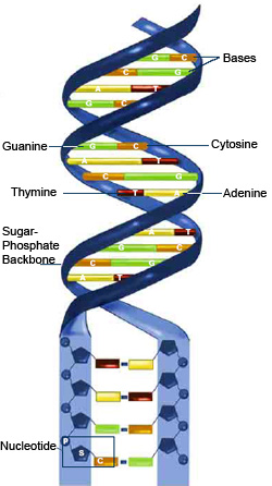
The diagram alongside illustrates the structure of the DNA, from the double helical structure shown at the top, to the more detailed structure towards the lower part of the diagram.
Note that the "backbone" is formed of sugar and phosphate, while the rungs are made of the Nitrogenous bases (Adenine, Thymine, Guanine, Cytosine) linked, with A pairing up with T, and G with C!
One sugar molecule, one phosphate molecule and a nitrogenous base together comprise a nucleotide. The DNA is therefore a chain of several such nucleotides.
Source: http://ahs-honorsbio2009-1.wikispaces.com/
Mitosis (Prophase, Metaphase & Anaphase of Mitosis)
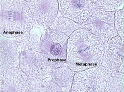
This photograph taken through a microscope shows several cells, one of them undergoing Anaphase of mitosis (observe the chromatids being pulled to the opposite poles), another in Metaphase (chromosomes are arranged along the equator)
Mitosis (Metaphase, Telophase)
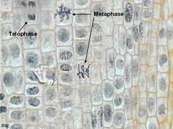
This image shows several cells as seen through a microscope. Note the different stages of cell division occurring in the different cells.
The Metaphase arrangement is clearly visible in two cells, where the chromosomes are arranged along the equator of the cell. Also, Telophase, where the chromatids have reached the opposite poles, and the rest of the cell is ready to divide.
The Metaphase arrangement is clearly visible in two cells, where the chromosomes are arranged along the equator of the cell. Also, Telophase, where the chromatids have reached the opposite poles, and the rest of the cell is ready to divide.
Meiosis
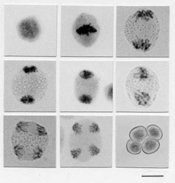
This sequence of photographs taken through a microscope illustrate meiosis occurring. Note that Meiosis I occurs through the first 6 images, and at the end of it, two cells with half the number of chromosomes are produced. In the last three images, the chromatids separate, and at the end, four cells are produced.
Note that meiosis began with a single cell having 2n chromosomes, and ended with 4 cells, each having n chromosomes!
Note that meiosis began with a single cell having 2n chromosomes, and ended with 4 cells, each having n chromosomes!
Click here to view more Resources on this and other topics.
